42 dissecting microscope diagram with labels
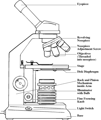
Microscope, Microscope Parts, Labeled Diagram, and Functions Stage with Stage Clips: The stage of a microscope is a flat platform where you place your subject slides. Stage clips hold the slides in place. The mechanical stage of your microscope will help you to move the slide around by turning two knobs. One knobs moves it left and right, the other knobs moves it up and down. Microscope Quiz: How Much You Know About Microscope Parts ... - ProProfs Projects light upwards through the diaphragm, the specimen, and the lenses. 5. Is used to regulates the amount of light on the specimen. Supports the slide being viewed. Moves the stage up and down for focusing. 6. Is used to support the microscope when carried. Moves the stage slightly to sharpen the image.
Cat Digestive System Anatomy with a Labeled Diagram The labeled diagram of the cat digestive system shows the following parts, structures, and organs - ... Microscope features of a cat stomach. The wall of a cat's stomach consists of mucosa, well-developed submucosa, a laminated tunica muscular layer, and a serosal covering. ... The details guide on cat dissection and organs from its ...

Dissecting microscope diagram with labels
Compound Microscope- Definition, Labeled Diagram, Principle, Parts, Uses In order to ascertain the total magnification when viewing an image with a compound light microscope, take the power of the objective lens which is at 4x, 10x or 40x and multiply it by the power of the eyepiece which is typically 10x. Therefore, a 10x eyepiece used with a 40X objective lens will produce a magnification of 400X. Microscope Under Cell Labeled Leaf Blank Microscope To Label microscope has an ocular objective of 10x and a high power objective of 50x what is the labelled diagram of a leaf cell under a microscope; ):Whole Mount (Entire Specimen or Organism) Autophagy plays an important role in plant-pathogen interactions - Buy this stock photo and explore similar images at Adobe Stock ... Simple Microscope - Parts, Functions, Diagram and Labelling Simple Microscope - Parts, Functions, Diagram and Labelling A microscope is one of the commonly used equipment in a laboratory setting. A microscope is an optical instrument used to magnify an image of a tiny object; objects that are not visible to the human eyes. Table of Contents The common types of microscopes are: What is a Simple microscope?
Dissecting microscope diagram with labels. Parts of a microscope with functions and labeled diagram Figure: Diagram of parts of a microscope There are three structural parts of the microscope i.e. head, base, and arm. Head - This is also known as the body. It carries the optical parts in the upper part of the microscope. Base - It acts as microscopes support. It also carries microscopic illuminators. Bright-field microscope (Compound light microscope) - Diagram (Parts ... Bright-field microscope parts (Labeled Diagram) Ocular Lens This microscope has two eye lenses or ocular lens on the top of the microscope that are used to focus the image from the objective lens. It is from these lenses that we see the magnified image of the specimen. Objective Lens Plant Cell Under Microscope 40X Labeled - Blogger Use tweezers to hold the frond, and use a dissecting needle to open sorus. Label the magnification under which the plant cells are being observed (40x or 100x). Examine a variety of cells with the compound microscope and estimate cell size. When carrying it, always use two hands, one on the base and one on the neck. Stereo Microscope - Parts, Types and Uses - Laboratoryinfo.com Stereo Microscope Parts and Functions It has three key parts namely: body, focus block, and viewing head/body. Let us take a look at the functions of every part. Body/viewing head - It houses the optical parts in the upper section of the microscope. Focus block - It attaches the head of the microscope to the stand and focuses it.
Microscope with labels afrikaans Microscope, Microscope Parts, Labeled Diagram, and. Bluetooth Label Printer for Autoclave. Microscope Labeling Worksheets - Learny Kids. Learning tools & flashcards, for free | Quizlet. Parts of a Microscope (Labeled) magnification. In a compound microscope the image from the objective lens is magnified again by the ocular lens. Parts of the Microscope with Labeling (also Free Printouts) Parts of the Microscope with Labeling (also Free Printouts) A microscope is one of the invaluable tools in the laboratory setting. It is used to observe things that cannot be seen by the naked eye. Table of Contents 1. Eyepiece 2. Body tube/Head 3. Turret/Nose piece 4. Objective lenses 5. Knobs (fine and coarse) 6. Stage and stage clips 7. Aperture dissecting microscope parts and functions pdf - Be Falcon Stereo microscope also known as Dissecting microscope is an optical instrument used for the observation of objects in low magnification, in which the instrument uses the light reflected from the surface rather than using the transmitted light from the object. • Relate the function of specific parts of Lumbricus to its locomotion. Microscopy- History, Classification, Terms, Diagram Microscopy can be defined as the scientific discipline of using microscopes for getting a magnified view of objects that can't be viewed by naked eyes. It is a very important tool in biology and nanotechnology. In microbiology, it is one of the most important tools used in observing microbial cells.
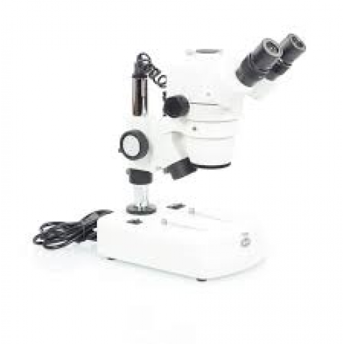
Welcome to microscopy4kids - Microscopy4kids Parts of Stereo Microscope (Dissecting microscope) - labeled diagram, functions, and how to use it A Stereo microscope is like a powerful magnifying glass, good for thick and solid specimens for observing the surface textures with 3D vision. Rs' Science 10 Everyday Things You Should Look at Under a Microscope Dissecting microscope (Stereo or stereoscopic microscope)- Definition ... Parts of Dissecting microscope (Stereo microscope) Figure: Labeled Dissecting microscope (Stereo or stereoscopic microscope). Image created using biorender.com LED illuminators- For some of the dissecting Microscopes, they have an inbuilt LED illuminator as a source of light. Dissecting microscope (Stereoscopic or Stereo microscope) This microscope is a dual-powered dissecting microscope of 10x-30x with an ability to rotate 360° making it ideal for viewing and focussing better to view samples. By rotating the lenses, users can change the magnification of image. Binocular Microscope Anatomy - Parts and Functions with a Labeled Diagram Now, I will discuss the details anatomy of the light compound microscope with the labeled diagram. Why it is called binocular: because it has two ocular lenses or an eyepiece on the head that attaches to the objective lens, this ocular lens magnifies the image produced by the objective lens. Binocular microscope parts and functions
Medical Technologists' Life in a Clinical Laboratory + Malaria Microscopy + Research ...
Difference Between Compound Microscope and Dissecting Microscope A Dissecting Microscope is also known as a stereomicroscope. It has a lower magnification power of 70x. A beam of light is projected above the specimen. This type of microscope is used to view larger specimens and to dissect small objects such as insects etc. It has a longer working distance of 150mm. Key Differences
Microscope Teaching Dissection
17 Parts of a Microscope (Optical and Structural Parts) It is a larger knob and is used to move the stage up or down very rapidly. The stage is raised or lowered rapidly with the help of a coarse adjustment knob. 14. Arm. Arm is a structural part of the microscope that connects the head (tube) with the base of the microscope. It provides support to the head.

Anatomy and Physiology I Coursework: Microscope A+P
Brightfield Microscope (Compound Light Microscope)- Definition ... Two focusing knobs i.e the fine adjustment knob and the coarse adjustment knob, found on the microscopes' arm, which can move the stage or the nosepiece to focus on the image. Their function is to ensure the production of a sharp image with clarity.
ScienCE BloG: #4 JournaL in SciencE
Scanning Electron Microscope (SEM) - Diagram, Working Principle ... Scanning electron microscope is a classification of electron microscope that uses raster scanning to produce the images of a specimen by scanning using a focused electron beam on the surface of the specimen. An SEM creates magnified images of the specimen by probing along a rectangular area of the specimen with a focused electron beam.
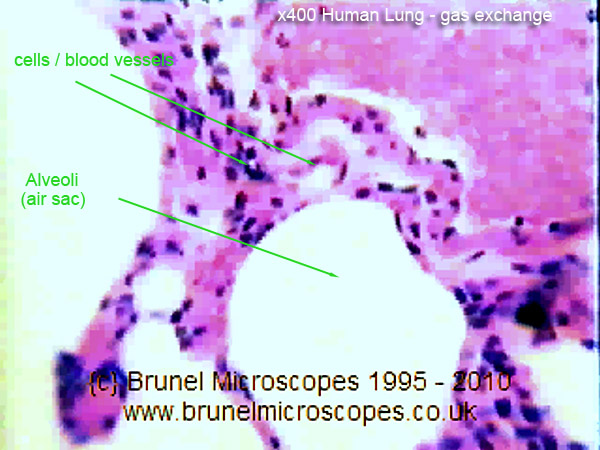
Human Cells - Part II an overview for light microscopists - Lungs
Simple Microscope - Diagram (Parts labelled), Principle, Formula and Uses A simple microscope consists of Optical parts Mechanical parts Labeled Diagram of simple microscope parts Optical parts The optical parts of a simple microscope include Lens Mirror Eyepiece Lens A simple microscope uses biconvex lens to magnify the image of a specimen under focus.

Compound Light Microscope Parts And Functions Worksheet | Decoratingspecial.com
Digital Microscope- Definition, Principle, Parts, Types, Examples, Uses Parts of a microscope with functions and labeled diagram Advantages of Digital Microscopes They provide high-resolution magnification of the images in pixels. It can tilt and provide 2D and 3D image measurements. They can store huge amounts of data, through imaging and recording movable and unmovable specimens
Post a Comment for "42 dissecting microscope diagram with labels"